IBL News Details
IBL News
Product News
- Product News2015/10/05
- Girdin Family Molecules: Girdin (pY1798, pY1764), Gipie, Daple Antibodies
 4 Girdin related antibodies have been relased. Girdin is a new protein playing an important role in the infiltration and metastasis of cancer cells. A new substance of Akt, Girdin (Girders of actin filament) was discovered by a research group led by Takahashi et al., Nagoya University by screening using yeast two-hybrid method and was elucidated that Girdin has an important role for cell migration downstream of Akt.
Girdin is phosphorylated on tyrosine 1798 when associated with structures required for migration. Omori K et al. Biochem Biophys Res Commun. 2015 Mar 20;458(4):934-40.
(New Products Released)
#28143 Anti-Human Girdin (Phospho-Y1798)Rabbit IgG A.P
#28145 Anti-Human Girdin (Phospho-Y1764)Rabbit IgG A.P
#28147 Anti-Human Daple Rabbit IgG Affinity Purify
#28149 Anti-Human Gipie Rabbit IgG Affinity Purify
Note: #28143 and #28145 are not available for sale in USA due to thrid parties' right.
#28143 Anti-Human Girdin (Phospho-Y1798) Rabbit IgG A.P. related images
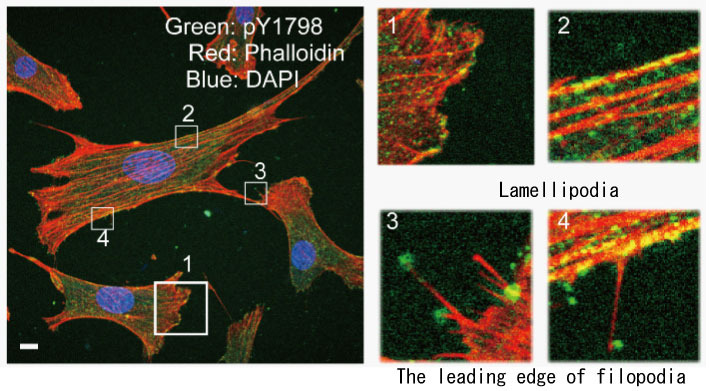 Distribution of phosphorylated Girdin in cultured NIH3T3 cells The cell was stained by phalloidin for visualizating of bundle of actin polymer. Point-like signal of pY1798 Girdin was colocalized with phalloidin positive stress fibers of NIH3T3 cells (2,4). The signal of pY1798 was recognized at lamelipodia (1) and the leading edge of filopodia (3,4).
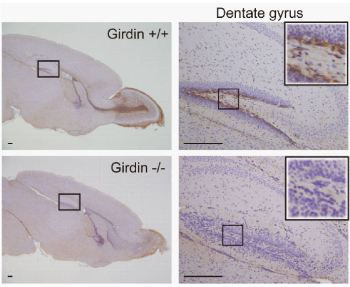 Distribution of pY1798 Girdin in mouse brain
Paraffin-embedded mouse brain tissue (14 days after birth): Stained Girdin native mouse (Girdin +/+), Girdin LacZ mouse knock-in model (Girdin -/-)using pY1798 Girdin antibody. Coloring by DAB and Contrast staining by hematoxylin. Left hand-side figures are low power and right hand-side figures are enlarged the part of the boxes. Positive signal was recognized in the hilum-side of the granule cell layer.
2 images above are provided by Prof. M. Takahashi, Department of Pathology, Nagoya University Graduate School of Medicine
(Daple, Gipie Antibody) Novel Girdin family molecules Wnt signal, for research of reticulum stress
Daple(Dishevelled-associating protein with a high frequency of leucine residues)
#28147 Anti-Human Daple Rabbit IgG Affinity Purify
Classical (β-catenin dependent) and non-classical (β-catenin non-dependent) Wnt signal pathway are signal pathways that regulate forming and maintaining of embryonic growth or cell structures of organs and amplifying of cells. It is also involved in various diseases including cancers. Especially, it has been well known that the non-classical Wnt signal pathway regulates polarity determination of tissues and migration of cells.
Daple is a molecule that was identified by A. Kikuchi et al, Osaka University and it shows that it has a homological sequence with Girdin or Gipie. It is suggested that Daple binds to Dvl and controls activation of Rac depended on Wnt5a stimulus via its mutual interaction. It was discovered that Daple is essential for cell migration and restructuring of actin structure and its molecular mechanism has also an important role for healing of wound of skin by Daple knockout mouse analysis
The Dishevelled-associating protein Daple controls the non-canonical Wnt/Rac pathway and cell motility. Ishida-Takagishi M et al. Nat Commun. 2012 May 29;3:859.
Gipie
#28149 Anti-Human Gipie Rabbit IgG Affinity Purify
A novel Girdin family molecule, Gipie is expressed in endothelial cells and it is induced by ER stresses. It interacts with GRP78 that is a 78kDa, glucose regulatory protein. It has been reported that Gipie controls IRE1-JNK signal pathway by the interactive function and protects endothelial cells from apoptosis that is induced by ER stresses under circumstances such as atherosclerosis or vascular endothelial failure.
Protective role of Gipie, a Girdin family protein, in endoplasmic reticulum stress responses in endothelial cells. Matsushita E et al. Mol Biol Cell. 2011 Mar 15;22(6):736-47.
Please feel free to contact us.
Immuno-Biological Laboratories Co., Ltd.
Diagnostic Research Reagent Division
Sales Support
TEL: +81-274-50-8666
Email: do-ibl@ibl-japan.co.jp