Product Detail
- HOME >
- For Researchers >
- Product Search >
- Search Result >
- #10385 Anti- Smad2L/3L (T220/T179 Phosphorylated) (1A1) Mouse IgG MoAb
Product Search
#10385 Anti- Smad2L/3L (T220/T179 Phosphorylated) (1A1) Mouse IgG MoAb

- Intended Use:
- Research reagents
- Application:
- WB, IP, IHC
- Package Size1:
- 50 μg
- Package Size2:
- 5 μg
- Note on Application Abbreviations
- WB:Western Blotting
- IP:Immunoprecipitation
- IHC:Immunohistochemistry
※ The product indicated as "Research reagents" in the column Intended Use cannot be used
for diagnostic nor any medical purpose.
※ The datasheet listed on this page is sample only. Please refer to the datasheet
enclosed in the product purchased before use.
Product Overview
Product Overview
| Product Code | 10385 |
|---|---|
| Product Name | Anti- Smad2L/3L (T220/T179 Phosphorylated) (1A1) Mouse IgG MoAb |
| Intended Use | Research reagents |
| Application | WB, IP, IHC |
| Species | Human |
| Immunizing antigen | Synthetic peptide of phosphorylated Smad2L/3L (T220/T179) |
| Source | Mouse-Mouse hybridoma (X63 - Ag 8.653 × BALB/c mouse spleen cells) |
| Clone Name | 1A1 |
| Subclass | IgG1 |
| Purification Method | Affinity purified with antigen peptide |
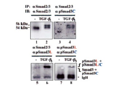
| Specificity | Reacts with phosphorylated Smad3L and Smad2L (Thr220/Thr179) of human in specific. |
| Package Form | Lyophilized product in PBS containing 1 % BSA and 0.05 % NaN3 |
| Storage Condition | 2 - 8℃ |
| Poisonous and Deleterious Substances | Applicable |
| Cartagena | Not Applicable |
| Package Size 1 | 50 μg |
| Package Size 2 | 5 μg |
| Remarks1 | The commercial use of products without our permission is prohibited. Please make sure to contact us and obtain permission. |
Product Description
Product Description
Phosphorylation of signal transduction molecules, Smad2 and Smad3, can be an important information for understanding of various biological functions of transforming growth factor (TGF)-β. TGF-β type I receptor and cyclin-dependent kinases phosphorylate both Smad2 and Smad3 at the C-terminals and at linker (middle) regions of them respectively. TGF-β signaling is mediated by Smad phosphoisoforms phophorylated at both the C-terminal and linker regions. This monoclonal antibody against for Smad2L/3L (T220/T179) recognizes the part of phosphorylated threonin (Thr220 of Smad2 and Thr179 of Smad3) located in linker region of Smad2/3 specifically. It can be used for western blotting, immunoprecipitation and immunohistochemistry. It is applicable for enzyme biochemical analysis of TGF-β signaling and makes it possible to visualize the intermolecular reaction of phosphorylated Smad signaling in human tissues and monitor them in real-time. Thus, analysis of TGF-β signaling with this antibody is expected to be widely applied to cancer serearch and fibrosis research, and to contribute to understanding the wide variety of life phenomina mediated by phosphoisoforms of Smad2/3.
Related Products
Related Products


Narrowed Search
Keyword Search